


PARISS® Hyperspectral Imaging Spectroscopy Instruments
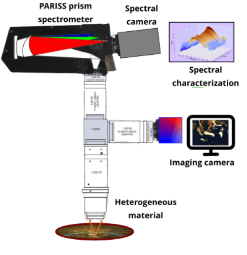
The PARISS imaging spectroscopy system
is modular and designed for laboratory-
based spectral and hyperspectral analysis
PARISS®: Prism And Reflector Imaging Spectroscopy System Basics
“Imaging Spectroscopy Requires a Highly Efficient Imaging Spectrometer”
The PARISS imaging spectroscopy system is based on a prism imaging spectrometer: Unlike a diffraction
grating that splits light into multiple diffraction orders, a prism delivers 90% of spectral intensity directly to
the detector. This ensures the highest possible signal to noise ratio (S/N). Go here for details.
Lightweight and compact, a PARISS system can be interfaced with a microscope for hyperspectral mi-
croscopy, or a variety of lens systems for application-specific spectral and hyperspectral imaging.
In order to increase instrument sensitivity, diffraction grating designs were discarded in favor of a prism-
based system.
The PARISS design was originally developed by The Aerospace Corporation for airborne remote sens-
ing. After redesign, the PARISS imaging spectrometer emerged as an ideal instrument for use in any
laboratory.
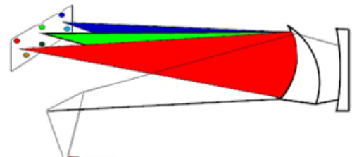
The curved sides of the prism add optical power to deliver near aberration-free imaging, from 365 up to
920-nm.
The net result is a highly significant improvement in signal to noise ratio (S/N) when compared to diffrac-
tion grating solutions. Enhanced sensitivity extends into the near IR where detectors and visible diffrac-
tion gratings are at their lowest efficiency.
The PARISS Prism: The Unique Curved Sides Dramatically Increases Sensitivity
Spectrum cameras: Located at the focus of the imaging spectrometer, the camera records the sample
spectra. Spectrum cameras are chosen as a function of anticipated signal strength.
LightForm partners with camera manufacturers that supply the appropriate camera consistent with an
application.
Zoom light-collection optics: LightForm integrates lenses optimized for the application. For example,
microscope objectives may be necessary for biopsy samples, while telephoto lenses would be used to
image ponds polluted with deadly cyanobacteria. There is no one lens solution that fits all applications.
LightForm specializes in developing zoom magnification optics that eliminate the need for multiple
lenses or objectives. While zoom camera lenses are commonplace most, if not all de-magnify a field of
view (FOV). PARISS zoom magnification optics accommodates objects that are less than one micron in
size.
Software: PARISS software is written in Python, with spectroscopy utilities that include: %reflection,
absorption, %transmission, fluorescence, luminescence… Math functions include smoothing,
background subtraction, division, multiplication, noise reduction…
Spectral classification: Spectra present that can be associated with objects or conditions are
assembled into classes. Selected classes can then be added to a “Reference Spectral Library.”
Correlation functions enable the extent of a class to be controlled and optimized.
Reference spectral libraries (RSL): RSLs consist of a collection of spectral classes consistent with a
given application or sample type. RSLs can be created, added to, or edited. Spectral classes can be
named and pseudo-colored
Spectra recognition algorithms: Enables objects in new samples to be “recognized” by correlating their
spectra with those in an RSL. A recognized object acquires the pseudo-color of the associated RSL
component class.


PARISS® Hyperspectral Imaging
Spectroscopy Instruments
The PARISS imaging spectroscopy system is
modular and designed for laboratory-based
spectral and hyperspectral analysis.
PARISS®: Prism And Reflector Imaging
Spectroscopy System Basics
The PARISS Prism: The Unique Curved
Sides Dramatically Increases Sensitivity
In order to increase instrument sensitivity,
diffraction grating designs were discarded in
favor of a prism-based system.
The PARISS design was originally
developed by The Aerospace Corporation
for airborne remote sensing.
After redesign, the PARISS imaging
spectrometer emerged as an ideal
instrument for use in any laboratory.
The curved sides of the prism add optical
power to deliver near aberration-free
imaging, from 365 up to 1,000-nm.
The net result is a highly significant
improvement in signal to noise ratio (S/N)
when compared to diffraction grating
solutions.
Enhanced sensitivity extends into the near
IR where detectors and visible diffraction
gratings are at their lowest efficiency.
Spectrum cameras: Located at the focus of
the imaging spectrometer, the camera
records the sample spectra. Spectrum
cameras are chosen as a function of
anticipated signal strength.
LightForm partners with camera
manufacturers that supply the appropriate
camera consistent with an application.
Zoom light-collection optics: LightForm
integrates lenses optimized for the
application.
For example, microscope objectives may be
necessary for biopsy samples, while
telephoto lenses would be used to image
ponds polluted with deadly cyanobacteria.
There is no one lens solution that fits all
applications.
LightForm specializes in developing zoom
magnification optics that eliminate the need
for multiple lenses or objectives.
While zoom camera lenses are
commonplace most, if not all de-magnify a
field of view (FOV).
PARISS zoom magnification optics
accommodates objects that are less than
one micron in size.
Software: PARISS software is written in
Python, with spectroscopy utilities that
include: %reflection, absorption,
%transmission, fluorescence,
luminescence…
Math functions include smoothing,
background subtraction, division,
multiplication, noise reduction…
Spectral classification: Spectra present
that can be associated with objects or
conditions are assembled into classes.
Selected classes can then be added to a
“Reference Spectral Library.”
Correlation functions enable the extent of a
class to be controlled and optimized.
Reference spectral libraries (RSL): RSLs
consist of a collection of spectral classes
consistent with a given application or sample
type.
RSLs can be created, added to, or edited.
Spectral classes can be named and pseudo-
colored
Spectra recognition algorithms: Enables
objects in new samples to be “recognized”
by correlating their spectra with those in an
RSL. A recognized object acquires the
pseudo-color of the associated RSL
component class.
Imaging Spectroscopy Requires a Highly
Efficient Imaging Spectrometer
The PARISS imaging spectroscopy system is
based on a prism imaging spectrometer:
Unlike a diffraction grating that splits light into
multiple diffraction orders, a prism delivers
90% of spectral intensity directly to the
detector.
This ensures the highest possible signal to
noise ratio (S/N).

